
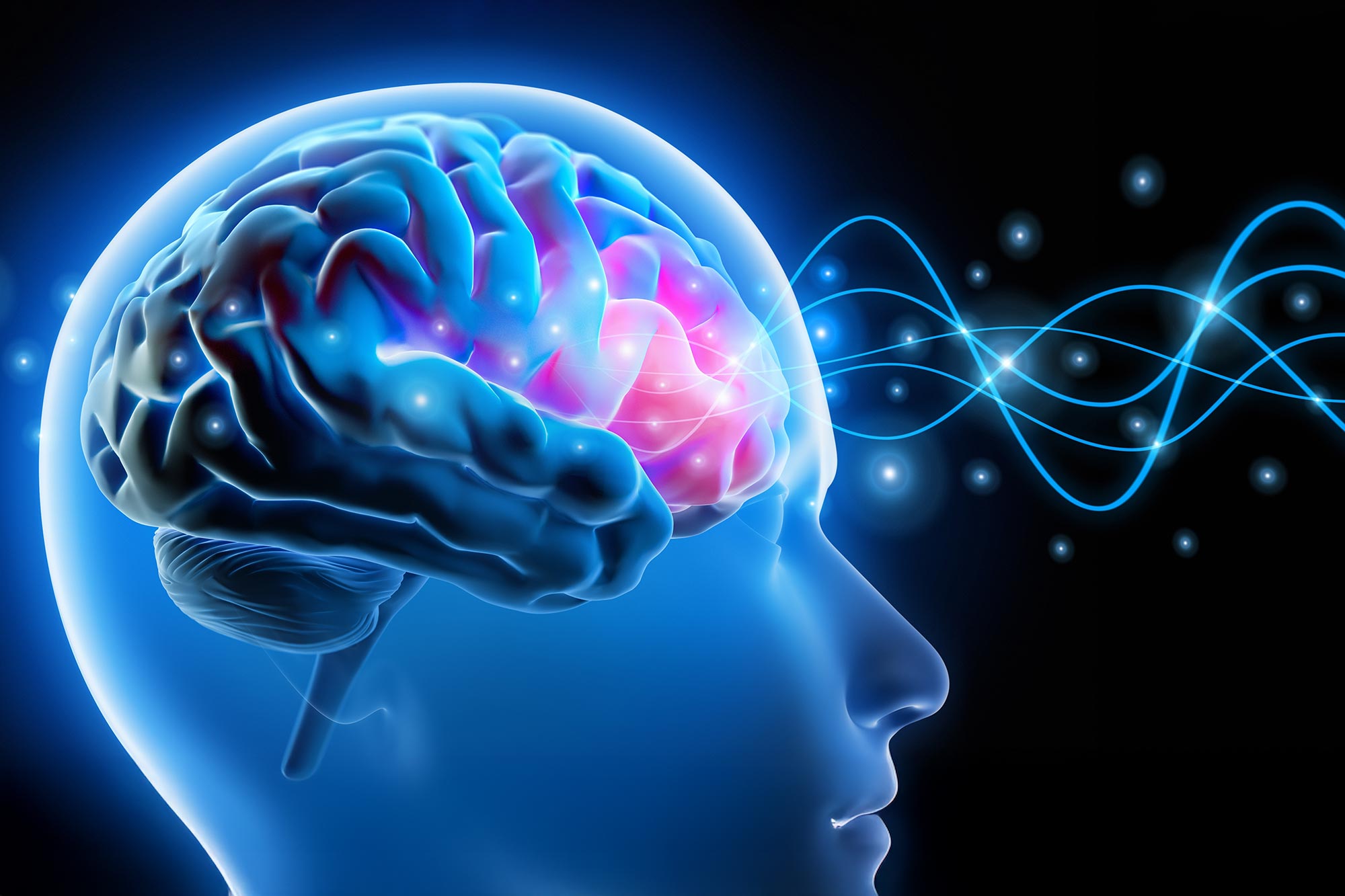
Scientists have visualized the brain’s hidden heartbeat, unlocking new clues to aging and Alzheimer’s.
Scientists at the Mark and Mary Stevens Neuroimaging and Informatics Institute (Stevens INI) at the Keck School of Medicine of USC have unveiled a breakthrough brain imaging method that captures how the brain’s tiniest blood vessels pulse with every heartbeat. These subtle rhythmic movements may provide new insight into how the brain ages and how diseases such as Alzheimer’s begin to develop.
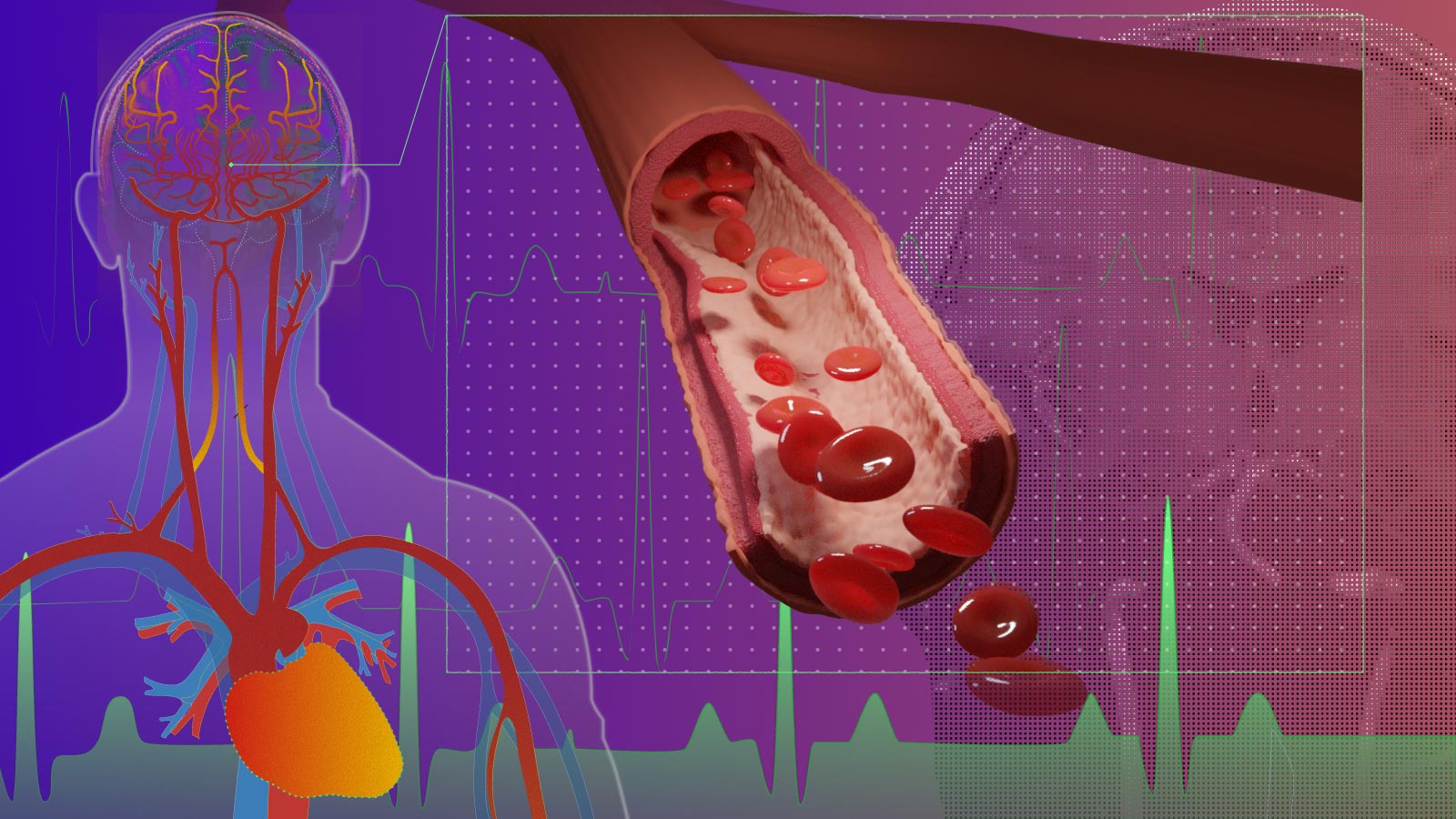
Published in Nature Cardiovascular Research, the study introduces the first noninvasive way to measure “microvascular volumetric pulsatility,” which refers to the rhythmic expansion and contraction of the brain’s smallest blood vessels in living people.
Using an ultra-high field 7T magnetic resonance imaging (MRI) scanner, the team discovered that these microvessel pulses become stronger with age, especially within the brain’s deep white matter—a region vital for communication between different brain networks. This area is particularly vulnerable as blood flow from the heart’s distal arteries weakens over time. Stronger pulsations in these vessels may disturb brain systems, potentially accelerating memory decline and contributing to Alzheimer’s disease.
The Brain’s Hidden Pulse and Its Role in Disease
“Arterial pulsation is like the brain’s natural pump, helping to move fluids and clear waste,” said Danny JJ Wang, PhD, professor of neurology and radiology at the Keck School of Medicine and senior author of the study. “Our new method allows us to see, for the first time in people, how the volumes of those tiny blood vessels change with aging and vascular risk factors. This opens new avenues for studying brain health, dementia, and small vessel disease.”
Researchers have long understood that stiffness and increased pulsation in large arteries are associated with stroke, dementia, and small vessel disease. However, until now, observing these same pulsations in the brain’s smallest vessels required invasive methods used only in animal studies.
Innovation in MRI: Seeing the Brain’s Smallest Rhythms
To solve this, the USC team merged two sophisticated MRI techniques, vascular space occupancy (VASO) and arterial spin labeling (ASL), to detect minute changes in microvessel volume over each heartbeat. Their results showed that older adults experience stronger microvascular pulsations in deep white matter than younger adults and that high blood pressure amplifies this effect.
“These findings provide a missing link between what we see in large vessel imaging and the microvascular damage we observe in aging and Alzheimer’s disease,” said lead author Fanhua Guo, PhD, a postdoctoral researcher in Wang’s lab.
Pulsations, Waste Clearance, and Alzheimer’s Risk
When these tiny blood vessel pulses become excessive, they may interfere with the brain’s “glymphatic system,” a recently identified network that clears away waste such as beta-amyloid, the protein that accumulates in Alzheimer’s disease. Disrupted fluid circulation over time could worsen cognitive decline.
“Being able to measure these tiny vascular pulses in vivo is a critical step forward,” said Arthur W. Toga, PhD, director of the Stevens INI. “This technology not only advances our understanding of brain aging but also holds promise for early diagnosis and monitoring of neurodegenerative disorders.”
The Next Frontier in Brain Health Research
The research team is now exploring how to adapt this method for wider use, including on more common 3T MRI scanners found in hospitals. Future work will investigate whether measuring microvascular volumetric pulsatility can predict changes in cognition and serve as an early biomarker for Alzheimer’s disease and related conditions.
“This is just the beginning,” Wang said. “Our goal is to bring this from research labs into clinical practice, where it could guide diagnosis, prevention, and treatment strategies for millions at risk of dementia.”
Reference: “Assessing cerebral microvascular volumetric with high-resolution 4D cerebral blood volume MRI at 7 T” by Fanhua Guo, Chenyang Zhao, Qinyang Shou, Ning Jin, Kay Jann, Xingfeng Shao and Danny JJ Wang, 25 September 2025, Nature Cardiovascular Research.
DOI: 10.1038/s44161-025-00722-1
In addition to Wang, the study’s other authors are Fanhua Guo, Chenyang Zhao, Qinyang Shou, Kay Jann, and Xingfeng Shao from the Stevens INI, and Ning Jin from Siemens Healthcare.
This research was supported by the National Institutes of Health (NIH) grants UF1-NS100614, S10-OD025312, R01-600 NS114382, R01-EB032169, RF1AG084072, R01-EB028297, R01-NS134712, and R01-NS121040.
Never miss a breakthrough: Join the SciTechDaily newsletter.
Follow us on Google, Discover, and News.


Discussion about this post